

First In-Vivo Real-Time Range-Guided Radiotherapy Using Radioactive Ion Beams
27.07.2026
In a study published in Communications Medicine, part of the Nature Portfolio, researchers from GSI/FAIR Darmstadt and LMU Munich have achieved the world’s first in-vivo demonstration of real-time, range-guided particle therapy using radioactive ion beams. The work was conducted within the EU-funded BARB project—Biomedical Applications of Radioactive Ion Beams.
By monitoring therapeutic carbon-11 (11C) ion beams in mice, the researchers demonstrated submillimetric beam-range verification, with updated in-beam PET images generated approximately every 25 seconds. The PET-guided beam positions correlated directly with biological outcomes: an undershooting beam resulted in continued tumor growth, whereas an overshooting beam achieved tumor coverage but caused toxicity in healthy tissue. The correctly positioned beam achieved tumor control while sparing the surrounding healthy organs.
Building on results from the SIRMIO project, the LMU team conceived and constructed the dedicated spherical in-beam PET scanner, developed the real-time image-reconstruction algorithms, and established the spatial co-registration framework required for dynamic treatment adaptation.
The following members of the LMU Chair of Medical Physics contributed to this collaborative effort, listed in their order of appearance in the publication: Francesco Evangelista and Munetaka Nitta, who are shared first authors together with GSI researchers Martina Moglioni and Tamara Vitacchio; Giulio Lovatti; Gabriele Corbetta; Jonathan Bortfeldt; and Peter G. Thirolf. Katia Parodi, BARB co-PI and associated partner at LMU, and Marco Durante, BARB PI at GSI, are shared senior authors.
This milestone highlights the potential of combining radioactive ion beams with real-time PET guidance to enable a new form of ultra-precise, adaptive particle therapy.
Link to publication: https://www.nature.com/articles/s43856-026-01786-1
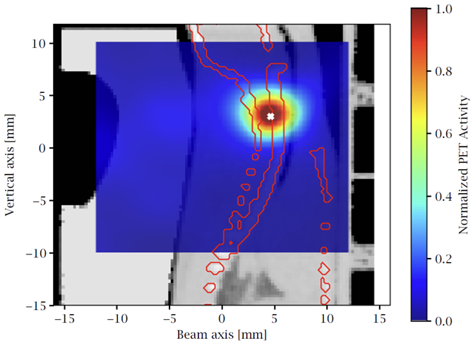
Figure: Online SIRMIO PET image of the positron activity distribution acquired during a probing beam 11C-irradiation prior to the entire dose delivery. The white “×” marker indicates the position along the beam axis corresponding to the 80% dose fall-off of the probing collimated 11C beam.