

WP2c PET Imaging
One of the solutions foreseen for in-vivo range verification in the SIRMIO platform, especially suited for application at continuous-wave cyclotrons and (slow-cycling) synchrotrons, is positron-emission-tomography (PET), for which an in-beam scanner has been designed to measure the irradiation-induced pattern of β+-activity, correlated to the primary proton beam range. Different detector technologies and geometrical arrangements have been simulated and investigated in terms of sensitivity and spatial resolution. Besides range verification, in-situ PET imaging could also open new opportunities for biological image guidance in the foreseen pre-clinical experiments. Hence, simultaneous integration of ionoacoustics/ultrasound and PET imaging is supported by the SIRMIO platform.
For the in-beam PET scanner, an optimum compromise has been sought within the given space constraints for compatibility with the SIRMIO beamline, the movable mouse holder and integration of US/ionoacoustics. The current design features a spherical-like assembly of 56 (partially tapered) LYSO scintillators with depth-of-interaction capability, according to detector technologies developed at the collaborating National Institute of Radiological Sciences, NIRS-QST, Japan. Iterative reconstruction of simulated positron point sources showed a promising resolution of 0.4 – 1.0 mm (FWHM) and detection efficiency from 12% to 7% in the relevant few (ca. ± 5) millimeters around the center of the scanner field of view, where the tumor will be located. Fig. 1 (left) illustrates the overall design of the spherical detector arrangement around the beam axis and vertical opening indicating the free space reserved for the mouse holder. 56 detector modules are arranged in 6 rings around the beam axis and the mouse holder, leaving enough open inner space (inner radius 72 mm) to accommodate also additional detectors (e.g. ultrasound transducers) and to allow for a longitudinal and transversal translation of the PET scanner by ca. 20 mm to enable placing the mouse tumor in the beam isocenter.
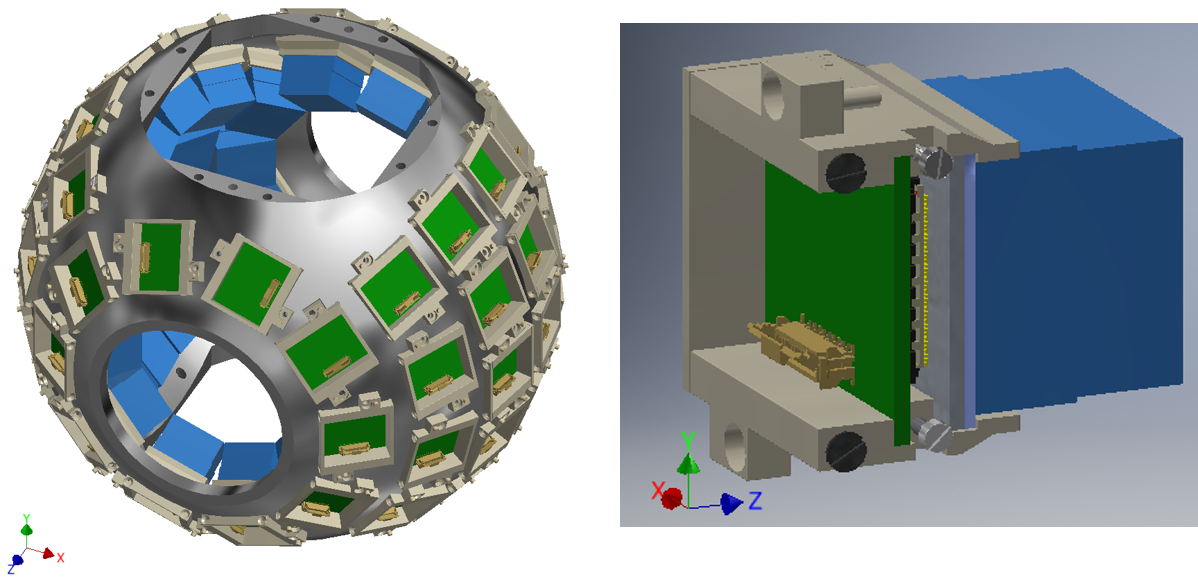
Fig. 1: Left: Schematic drawing of the dedicated in-beam PET scanner design for SIRMIO, with the opening for the mouse holder in the vertical direction, while the beam enters horizontally. The 56 detector blocks are mounted to a spherical support frame. Right: Schematical view of a DOI PET detector block with a tapering in x-y direction and its related SiPM photosensor array and frontend signal processing board mounted into a support frame that will connect to the spherical frame of the left panel.
Fig. 1 (right) illustrates the setup of an individual detector block, here showing one of the options under study consisting of a block of LYSO crystal pixels, arranged in a 3-layer DOI geometry. Also shown is the (8x8) SiPM photosensor array and (in green) the charge division circuit (CDC) that reduces the 64 signal channels to 4 by applying an Anger logic.
Contact
PD Dr. Peter G. Thirolf
Dr. Munetaka Nitta
People
Giulio Lovatti, Dr. Munetaka Nitta, Prof. Katia Parodi, Dr. Peter G. Thirolf, Dr. Mohammad Safari, Dr. George Dedes, Roghieh Haghani
References
K. Parodi, W. Assmann, C. Belka, J. Bortfeldt, D.-A. Clevert, G. Dedes, R. Kalunga, S. Kundel, N. Kurichiyanil, P. Lämmer, J. Lascaud, K. Lauber, G. Lovatti, S. Meyer, M. Nitta, M. Pinto, M. Safari, K. Schnürle, J. Schreiber, P.G. Thirolf, H.-P. Wieser, M. Würl, Towards a novel small animal proton irradiation platform – the SIRMIO project, Acta Oncologica (2019), https://doi.org/10.1080/0284186X.2019.1630752
M. Nitta, G. Lovatti, G. Dedes, S. Takyu, M. Safari, T. Binder, G. Vinci, T. Yamaya, K. Parodi, P.G. Thirolf, Investigation of the dependency of properties of a 4-layer DOI PET detector on the scintillator material, Poster presented at the DPG Spring Meeting, Regensburg, 2019, Verhandl. DPG (VI), 54, ST 4.6 (2019)
G. Lovatti, M. Nitta, M. Safari, J. Bortfeldt, A. Zoglauer, P.G. Thirolf, G. Dedes, and K. Parodi, Design study of a novel geometrical arrangement for an in-beam small animal PET scanner, IEEE NSS/MIC M-06-182, Manchester (UK), 28.10-1.11.2019.
M. Nitta, G. Lovatti, M. Safari, G. Dedes, T. Yamaya, P. G. Thirolf, K. Parodi, Sensitivity comparison of LYSO and GAGG scintillators for In-beam PET Measurements, IEEE NSS/MIC, M-06-365, Manchester (UK), 28.10-1.11.2019
M. Nitta, G. Lovatti, M. Safari, G. Dedes, H. Tashima, T. Yamaya, P. G. Thirolf, K. Parodi, Simulation Study of a Pyramid-like 4-layer DOI Detector with the Sparse Reflector Insertion, IEEE NSS/MIC, M-06-079, Manchester (UK), 28.10-1.11.2019