Image Guidance
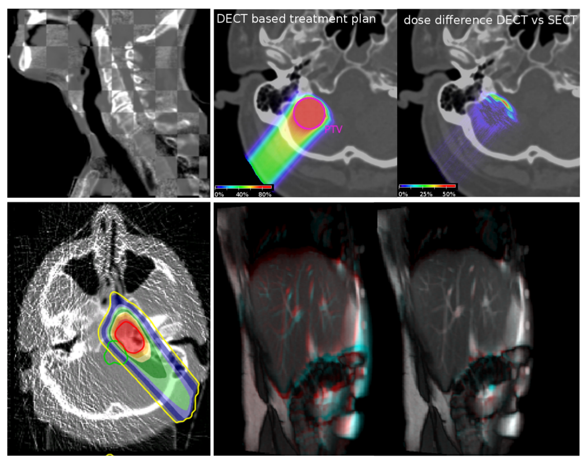
Image Guided Radiation Therapy (IGRT) refers to the systematic use of imaging throughout the treatment course in external beam radiotherapy. The application of image guidance is meant to reduce uncertainties in the localization of tumors and organs at risk at the time of irradiation, thus maximizing the potential of highly conformal treatments. In case accelerated ion beams are used, the role of image guidance is even more important, due to their enhanced selectivity for dose deposition and the resulting higher sensitivity to uncertainties. The availability of accurate imaging at the time of treatment empowers the implementation of adaptive treatment protocols, where treatment plans are evaluated and re-optimized as a function of the measured anatomical changes or, in case of ions, improved knowledge of the tissue stopping properties. We are currently pursuing several research projects in the field of IGRT, including cone-beam computed tomography (CBCT), dual-energy CT (DECT), proton and ion CT and Magnetic Resonance Imaging (MRI).
-
Our research on cone beam computed tomography (CBCT) focusses on its application to image-guided proton therapy. Proton therapy has lagged conventional linac-based photon therapy, where volumetric CBCT imaging is widespread, in part due to the limited amount and bespoke nature of proton therapy facilities. However, the technology has been recently rapidly adopted and several centers now rely on CBCT imaging for patient positioning.
more
-
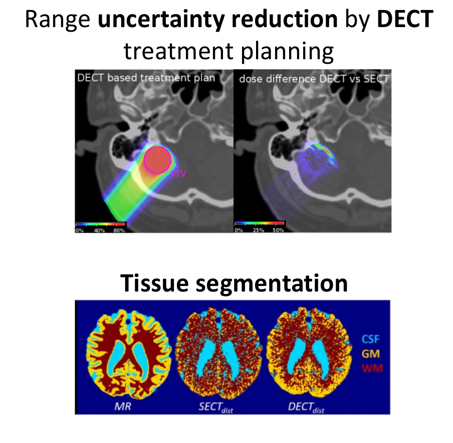
By acquiring CT images at different photon energies, DECT allows medical physicists to deduce material properties which influence the linear attenuation coefficient. This has clear benefits for proton therapy, where the proton range in tissues is strongly correlated to the electron density, which is one of the quantities DECT excels at measuring.
more
-
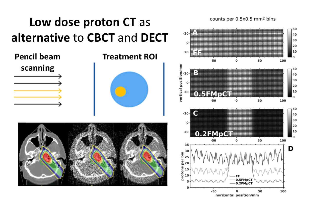
While DECT shows promising results for conversion of linear attenuation coefficients to proton (ion) stopping power, proton (ion) CT bypasses conversion entirely by imaging the stopping power directly. Proton/ion CT relies on measurements of the beam energy downstream from the patient, with optional particle-by-particle tracking technology for improved resolution. The LMU team is part of or collaborates with several groups developing proton or heavier ion CT scanners.
more
-
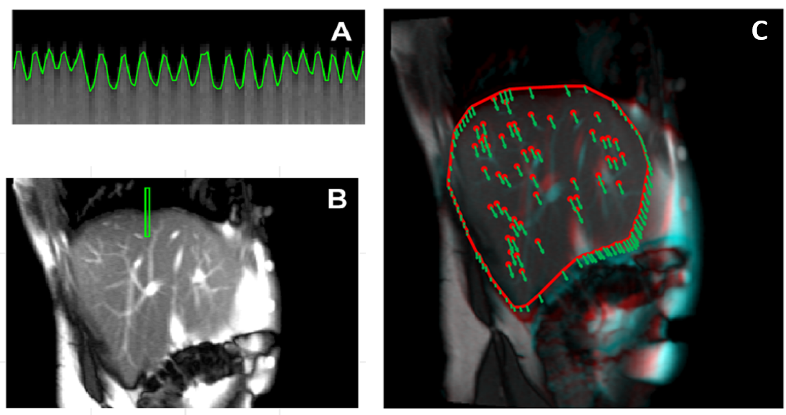
The interest toward Magnetic Resonance Imaging (MRI) in radiation oncology is rapidly increasing, mainly due to its capability of imaging without ionizing radiation at superior soft tissue contrast, compared to conventional X-ray imaging. Technological developments are currently ongoing to integrate MRI in the external-beam radiotherapy workflow, including imaging for guidance at the linear accelerator.
more