

Cone Beam CT
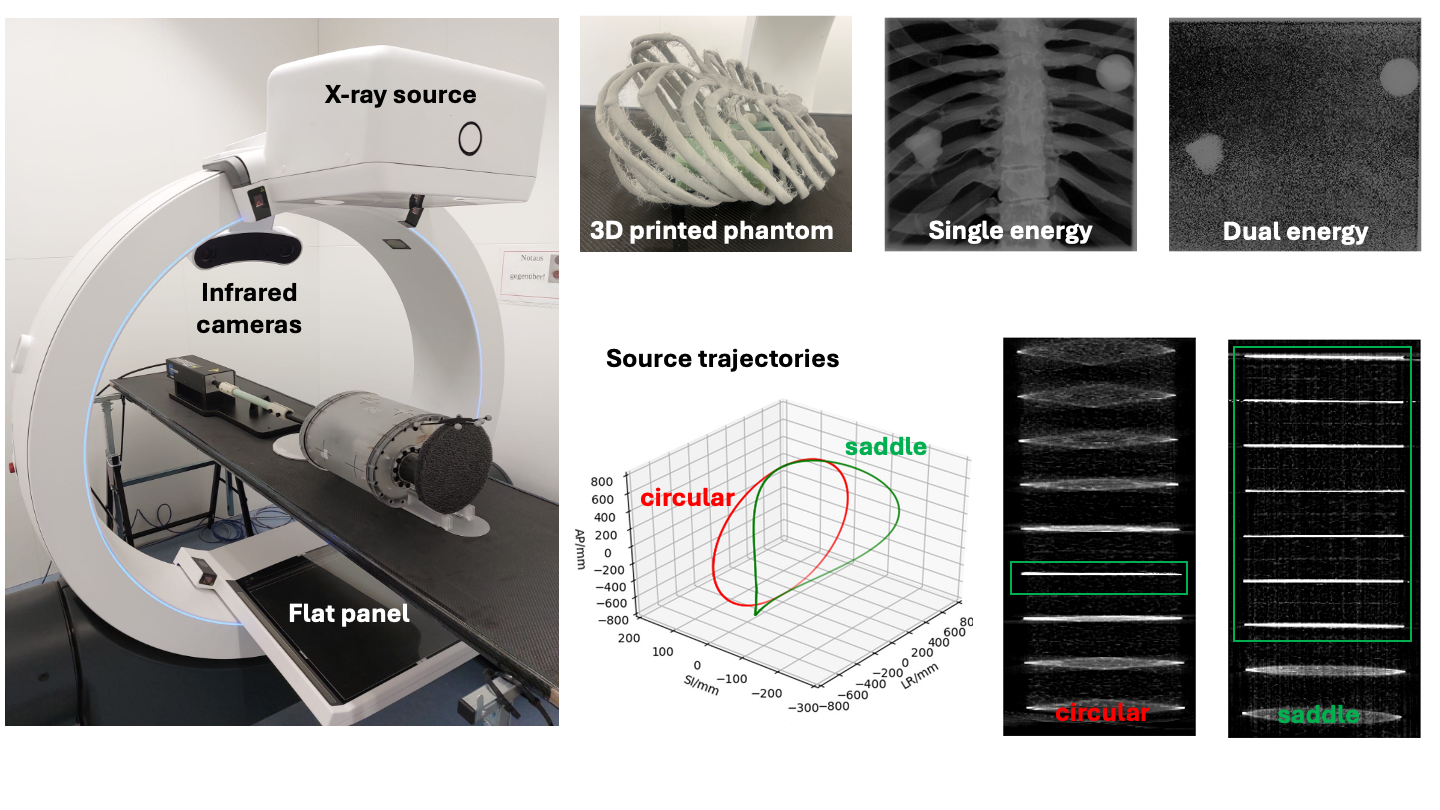 Our research on cone beam computed tomography (CBCT) is focused on advancing current imaging methods to improve the soft tissue contrast and reduce reconstruction artifacts. Examples include (i) the implementation of dual-energy imaging protocols and (ii) the use of non-circular CBCT trajectories. In dual-energy mode pulsed X-ray imaging is applied by switching the energy in between consecutive pulses. This provides a method to differentiate between different tissues, e.g. to separate the attenuation due to bone vs. soft tissue. Considering volumetric reconstruction, circular trajectories in CBCT imaging produce reconstruction artifacts when moving away from the plane of the source trajectory. We therefore exploit the use of non-circular CBCT trajectories to enlarge the area where artifact-free reconstruction can be achieved.
Experimental activities for CBCT-based research rely on our robotic CBCT scanner (ImagingRing Mobile, medPhoton), financed by the Deutsche Forschungsgemeinschaft (DFG) under the Major Research Instrumentation program. Access to the device is possible for external groups also, by contacting the responsible person (Prof. Dr. Marco Riboldi)
Contact:
Prof. Dr. Marco Riboldi
References:
Albrecht J, Rit S, Steininger P, Ginzinger F, Huber P, Messner I, Kraihamer M, Schmitz H, Corradini S, Belka C, Kurz C, Riboldi M, Landry G. Cone-beam CT from complete data using saddle trajectories on a mobile robotic CBCT scanner. Medical Physics (2022) https://doi.org/10.1002/mp.16943
Wei C, Albrecht J, Rit S, Laurendeau M, Thummerer A, Corradini S, Belka C, Steininger P, Ginzinger F, Kurz C, Riboldi M, Landry G. Reduction of cone-beam CT artifacts in a robotic CBCT device using saddle trajectories with integrated infrared tracking. Medical Physics (2024) doi: 10.1002/mp.16943
Wei C, Rit S, Laurendeau M, Steininger P, Ginzinger F, Riboldi M, C Kurz, Landry G. Comparison of saddle and helical trajectories of a mobile cone beam computed tomography scanner with online calibration correction. Proceedings of the XXth International Conference on the use of Computers in Radiation therapy (2024) https://hal.science/hal-04753338/document
Alqethami N, Xie W, Palaniappan P, Ginzinger F, Steininger P, Riboldi M. 2247: Validation of an integrated platform for markerless motion tracking using dual energy X-ray imaging. Radiotherapy and Oncology (2024) https://doi.org/10.1016/S0167-8140(24)02484-8
Wei C, Albrecht J, Rit S, Laurendeau M, Thummerer A, Corradini S, Belka C, Steininger P, Ginzinger F, Kurz C, Riboldi M, Landry G. Reduction of cone-beam CT artifacts in a robotic CBCT device using saddle trajectories with integrated infrared tracking. Medical Physics (2024) https://doi.org/10.1002/mp.16943
Alqethami N, Xie W, Palaniappan P, Ginzinger F, Steininger P, Riboldi M. 1364 Motion detection accuracy of dual-energy X-ray imaging for lung cancer tracking: an experimental phantom study. Radiotherapy and Oncology (2025) https://doi.org/10.1016/S0167-8140(25)00412-8
Currently funded projects:
INSIGHT "Intelligent Tumor Tracking for Radiotherapy” (INSIGHT), funded by the German Federal Ministry of Research, Technology and Space (BMFTR) in the funding initiative "New Therapy Options through Innovative Medical Technology" (project number 13GW0730C). The project is in cooperation with Brainlab, the LMU Klinikum Radiation Oncology Department and the Radiation Oncology Department of Uniklinikum Erlangen.
DFG Image-guided brachytherapy with real-time navigated needle insertion (DFG project number 562329381). The project is in cooperation with the LMU Klinikum Radiation Oncology Department and the Radiation Oncology Department of Uniklinikum Erlangen.