

Magnetic Resonance Imaging
The interest toward Magnetic Resonance Imaging (MRI) in radiation oncology is rapidly increasing, mainly due to its capability of imaging without ionizing radiation at superior soft tissue contrast, compared to conventional X-ray imaging. Technological developments are currently ongoing to integrate MRI in the external-beam radiotherapy workflow, including imaging for guidance at the linear accelerator.
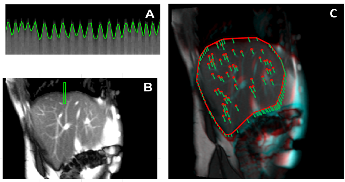
Research on MRI-guided radiotherapy at the Chair of Medical Physics is specifically focused on the use of MRI for treatment planning and 4D MRI guidance for tumors susceptible to significant breathing motion. Current research activities pertain MRI-based motion quantification for treatment planning and delivery, including 4D MRI sorting and real-time motion tracking in cine-MR images, as depicted below. We are developing 4D motion models to enable full 3D motion detection during treatment relying on cine-MR and 4D treatment planning information. Specific efforts are also dedicated to the implementation and testing of computational phantoms, aiming at quantitative validation of 4D MRI-guided strategies.
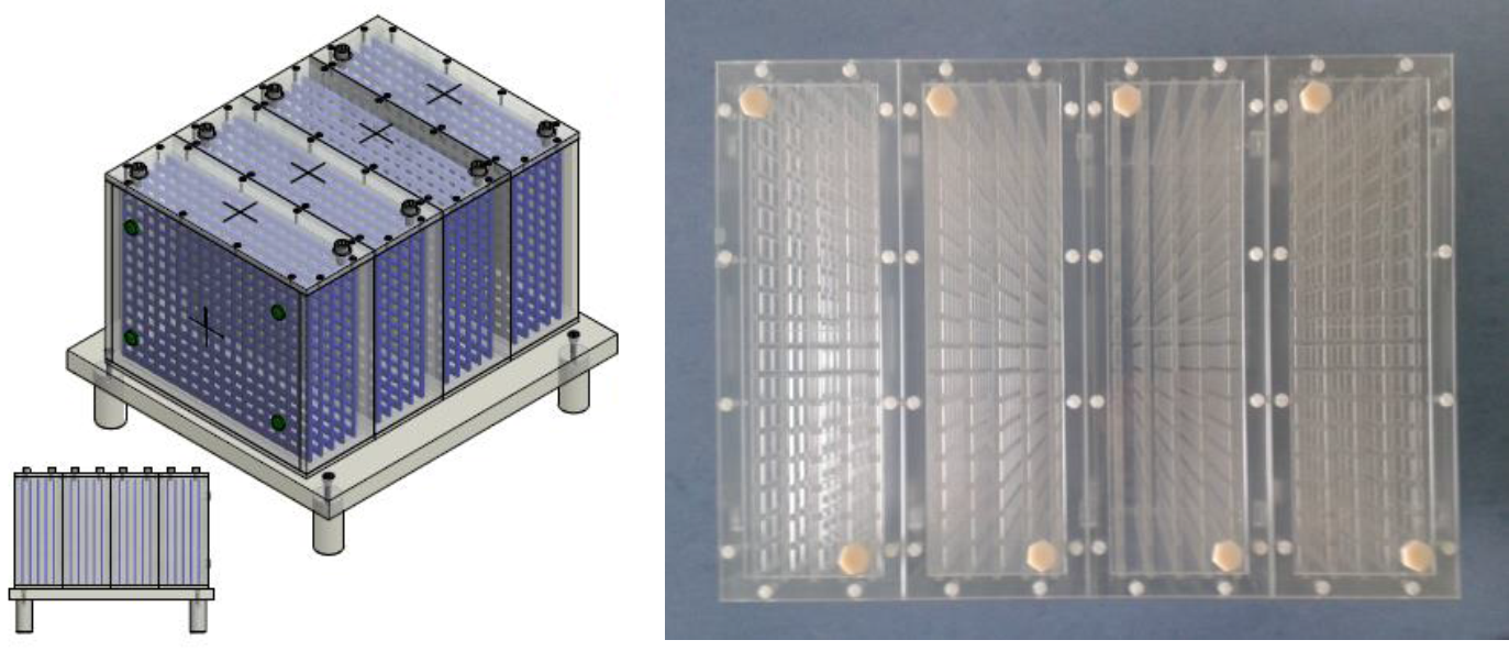
A key issue in motion management based on MRI guidance is the ability to handle MRI spatial distortions, as induced by static field inhomogeneity, non-linearity of the applied field gradients and patient dependent susceptibility effects. We are working on a comprehensive approach to spatial distortion modeling, including experimental measurements, numerical simulations, and dedicated phantom developments (see figure below).
Finally, the application of MRI guidance to proton therapy is currently being explored, focusing specifically on MR-based measurement methods to visualize proton beam delivery. We pursue numerical simulations and innovative experimental setup at ultra-low field based on optical methods, aiming at the maximal sensitivity in detecting the proton beam current and magnetization.
Contact:
Prof. Dr. Marco Riboldi
References:
Paganelli C, Kipritidis J, Lee D, Baroni G, Keall P, Riboldi M. Image-based retrospective 4D MRI in external beam radiotherapy: A comparative study with a digital phantom. Med Phys. 2018 May 14. doi: 10.1002/mp.12965. [Epub ahead of print]
Paganelli C, Lee D, Kipritidis J, Whelan B, Greer PB, Baroni G, Riboldi M, Keall P. Feasibility study on 3D image reconstruction from 2D orthogonal cine-MRI for MRI-guided radiotherapy. J Med Imaging Radiat Oncol. 2018 Feb 11. doi: 10.1111/1754-9485.12713. [Epub ahead of print]
Seregni M, Paganelli C, Summers P, Bellomi M, Baroni G, Riboldi M. A Hybrid Image Registration and Matching Framework for Real-Time Motion Tracking in MRI-Guided Radiotherapy. IEEE Trans Biomed Eng. 2018 Jan;65(1):131-139. doi: 10.1109/TBME.2017.2696361.
Paganelli C, Summers P, Gianoli C, Bellomi M, Baroni G, Riboldi M. A tool for validating MRI-guided strategies: a digital breathing CT/MRI phantom of the abdominal site. Med Biol Eng Comput. 2017 Nov;55(11):2001-2014
Seregni M, Paganelli C, Lee D, Greer PB, Baroni G, Keall PJ, Riboldi M. Motion prediction in MRI-guided radiotherapy based on interleaved orthogonal cine-MRI. Phys Med Biol. 2016 Jan 21;61(2):872-87. doi: 10.1088/0031-9155/61/2/872
Paganelli C, Lee D, Greer PB, Baroni G, Riboldi M, Keall P. Quantification of lung tumor rotation with automated landmark extraction using orthogonal cine MRI images. Phys Med Biol. 2015 Sep 21;60(18):7165-78. doi: 10.1088/0031-9155/60/18/7165
Paganelli C, Summers P, Bellomi M, Baroni G, Riboldi M. Liver 4DMRI: A retrospective image-based sorting method. Med Phys. 2015 Aug;42(8):4814-21. doi: 10.1118/1.4927252.
Paganelli C, Seregni M, Fattori G, Summers P, Bellomi M, Baroni G, Riboldi M. Magnetic resonance imaging-guided versus surrogate-based motion tracking in liver radiation therapy: a prospective comparative study. Int J Radiat Oncol Biol Phys. 2015 Mar 15;91(4):840-8. doi: 10.1016/j.ijrobp.2014.12.013.