Range Verification in Particle Therapy
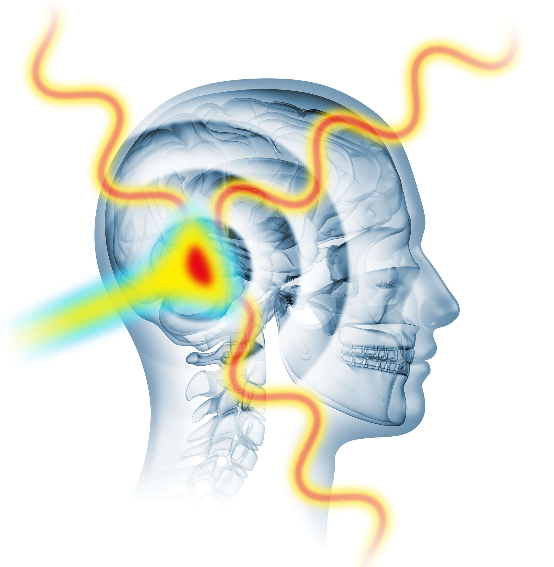
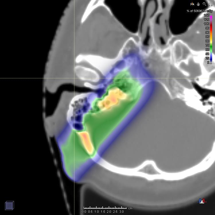
Particle therapy (PT) with protons and heavier ion beams is rapidly emerging as a promising radiation therapy modality due to the superior ability to concentrate energy deposition in the tumor, while better sparing normal tissue and critical organs compared to widely established photons. Despite considerable recent technological advances, particularly regarding ion accelerators (see also research area Laser Ion Acceleration”) and scanning beam delivery systems, along with the integration of in-room volumetric X-ray imaging (see research area Image guidance), full clinical exploitation of the favorable ballistic properties of ion beams is still hampered by the yet unsolved problem of range uncertainties in tissue. Although these uncertainties can be mitigated by the introduction of cautious safety margins and careful choice of beam directions, new approaches are being extensively investigated to tackle this issue at the stage of treatment planning (see research areas Image guidance and Dosimetry) or treatment delivery. For the latter task, we are currently investigating unconventional imaging modalities for in vivo verification of the beam range and thus localization of the maximum dose deposition (see figure), exploiting online/post-treatment detection of acoustic and nuclear-based emissions induced by ion interaction in tissue.
-
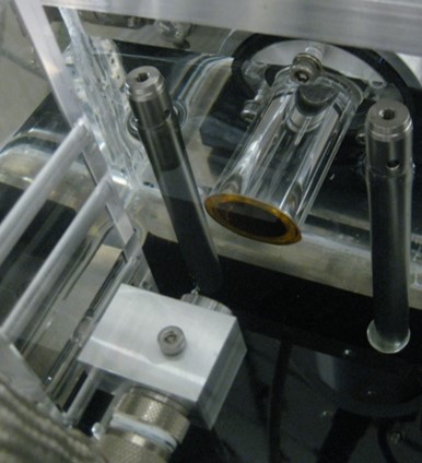
When penetrating a medium, ions mainly lose energy in electronic collisions, resulting in localized heating and a thermal expansion, which generates thermoacoustic emissions detectable with acoustic transducers. Being the thermoacoustic emission naturally enhanced at the maximum of ion energy deposition (so-called Bragg-peak), time-of-flight (TOF) measurements in combination with knowledge of the speed of sound in the traversed medium can enable recovery of the ion range. Especially in combination with morphological ultrasonography, this so-called “ionoacoustics” could offer a compact and cost-effective modality for real-time in-vivo verification of the beam range, co-registered to tissue anatomy, at least for suitable indications of feasible sonic access.
more
-
One class of secondary emissions originating from the interaction of a therapeutic ion beam with tissue features prompt photons, produced in fast (~ns) de-excitation of excited nuclei following nuclear interactions between the particle beam and the organic tissue components (predominantly O,C,N). These photons span a multi-MeV energy range between ca. 3 MeV and 6 MeV.
more
-
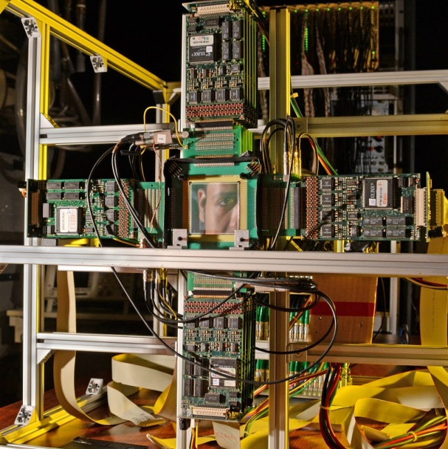
Among the clinically most investigated techniques for monitoring proton or heavier ion treatment is positron emission tomography (PET), which relies on the detection of coincident 511 keV gamma pairs produced in the annihilation of positrons released in the decay of β+-emitters produced in nuclear interactions between the incoming ions and the traversed tissue.
more
-
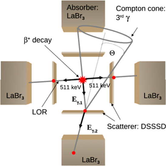
So far a whole class of potential PET isotopes, where in addition to the two back-to-back emitted 511 keV annihilation photons also a third, high-energy γ ray is emitted from an excited state in the daughter nucleus, has been excluded from medical application. The resulting extra dose delivered to the patient, as well as the expected increase of background from Compton scattering or even pair production, prevented the use of isotopes such as 44mSc, 86Y, 94Tc, 94mTc, 152Tb, or 34mCl. However, provided the availability of customized gamma cameras, this alleged disadvantage could be turned into a promising benefit, offering a higher sensitivity for the reconstruction of the radioactivity distribution in PET examinations.
more