

Dual Energy CT
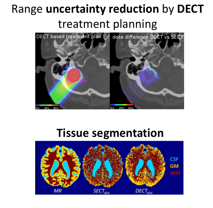
By acquiring CT images at different photon energies, DECT allows medical physicists to deduce material properties which influence the linear attenuation coefficient. This has clear benefits for proton therapy, where the proton range in tissues is strongly correlated to the electron density, which is one of the quantities DECT excels at measuring. DECT research at the LMU, performed in close collaboration with our colleagues from MAASTRO Clinic and the LMU Radiology Department, has focused on two broad applications: 1) the use of DECT to improve proton dose calculation accuracy by improved proton stopping power estimation and 2) exploiting the capability of DECT for better estimating the composition of human tissues. The second application has important implications for proton therapy, where range monitoring via nuclear-based secondary particle emission, which is highly tissue dependent, is an important area of research. By better estimating tissue composition from DECT, medical physicists are thus able to better predict the signal from secondary radiation and match it to what is observed in their detectors (see research on PET and prompt gamma).
We are currently actively combining DECT imaging with Monte Carlo simulations for dose calculation and secondary radiation prediction, as well as devising novel methods to validate the improvements of DECT using biological tissues. These activities are part of project B4 of the DFG funded Graduiertenkolleg GRK 2274.
Contact:
Dr. G. Landry
Prof. Dr. K. Parodi
References:
I.P. Almeida, L.E. Schyns, A. Vaniqui, B. van der Heyden, G. Dedes, A.F. Resch, F. Kamp, J.D. Zindler, K. Parodi, G. Landry*, F. Verhaegen*. Monte Carlo proton dose calculations using a radiotherapy specific dual-energy CT scanner for tissue segmentation and range assessment. Phys Med Biol (2018).
*Authors contributed equally
I.P. Almeida*, G. Landry*, G. Dedes, R. Patel, M. Pankuch, G. Coutrakon, R Schulte, F. Verhaegen, K. Parodi, J.C. Roeske. Evaluating clinical stopping power estimation from a radiotherapy dual energy CT scanner. Acta Phys Pol B (2017) vol 48 (10) pp. 1619
*Authors contributed equally
B. Bernt*, G. Landry*, F. Schwarz, T. Tessonnier, F. Kamp, G. Dedes, C. Thieke, M. Wuerl, C. Kurz, U. Ganswindt, F. Verhaegen, J. Debus, C. Belka, W. Sommer, M. Reiser, J. Bauer, K. Parodi. Application of single- and dual-energy CT brain tissue segmentation to PET monitoring of proton therapy. Phys Med Biol (2017) vol. 62 (6) pp. 2427
*Authors contributed equally
N. Hudobivnik, F. Schwarz, T. Johnson, L. Agolli, G. Dedes, T. Tessonnier, F. Verhaegen, C. Thieke, C. Belka, W. Sommers, K. Parodi, G. Landry. Comparison of proton therapy treatment planning for head tumors with a pencil beam algorithm on dual and single energy CT images. Med Phys (2016) vol. 43 (1) pp. 495-504
D. Hansen, J. Seco, T. Sangild, T.S. Sorensen, J.B.B. Petersen, J.E. Wildberger, F. Verhaegen, G. Landry. A simulation study on proton CT stopping power accuracy using dual energy CT scans as benchmark. Acta Oncol (2015) vol. 54 (9) pp. 1638-1642
Currently funded projects: