

Proton/Ion CT
While DECT shows promising results for conversion of linear attenuation coefficients to proton (ion) stopping power, proton (ion) CT bypasses conversion entirely by imaging the stopping power directly. Proton/ion CT relies on measurements of the beam energy downstream from the patient, with optional particle-by-particle tracking technology for improved resolution. The LMU team is part of or collaborates with several groups developing proton or heavier ion CT scanners.
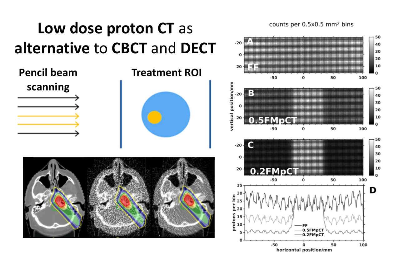
Besides high stopping power accuracy, proton/ion CT offers the prospect of frequent image guidance at the isocenter, enabling both image-guided and adaptive particle therapy. The modality is particularly appealing due to reportedly better contrast to noise ratio at equivalent dose when compared to X-ray CT imaging. To enable an even further reduction of the imaging dose we introduced the concept of fluence modulated proton computed tomography (FMpCT), adopting the ideas of fluence field modulation from X-ray CT. This can be achieved by modulating the proton fluence within a projection, allowing the eventual prescription of image quality in relevant portions of the image, such as the proton beam path. Fluence modulation within a projection is readily achievable by using the pencil beam scanning (PBS) capability of modern proton therapy beamlines. Image quality prescription can be performed either in a “forward planning” fashion, where high fluence is preserved for pencil beams intersecting a region of interest (ROI), or in an “inverse planning” fashion where a model linking image quality and proton pencil beam (PB) fluence is required for optimization.
At the LMU, in the context of the DFG funded project on FMpCT (grant # 388731804), we investigate proof of principle simulation studies, experimental FMpCT scans on the basis of PBS and finally the fundamentals of noise reconstruction in pCT. We work closely with our colleagues from the proton CT collaboration (Loma Linda University / University of California Santa Cruz / Northwestern Chicago Proton Therapy Center) and reconstruction experts from CREATIS in Lyon.
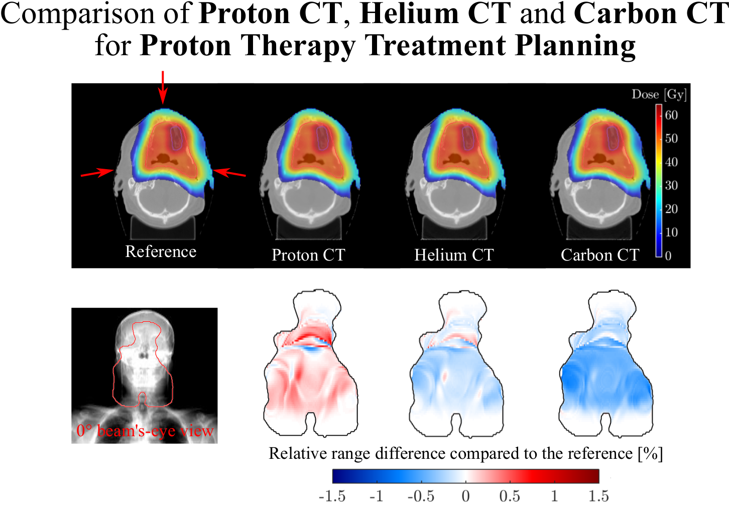
In the framework of the DFG-funded project “Hybrid ImaGing framework in Hadrontherapy for Adaptive Radiation Therapy (High-ART)” (grant # 372393016), we are evaluating the imaging performance of different ion species and studying the imaging capabilities of different detector modalities based both on particle-by-particle tracking as well as on the integrated Bragg peak signals of all the ions of the pencil beam. To this end, we rely on computational imaging frameworks, spanning from dedicated (fast) analytical simulators to more complex Monte Carlo simulations, also embedding radiobiological models in cooperation with collaborators from Yale School of Medicine. We apply specific data processing to the Bragg peak signal and we investigate new tomographic image reconstruction algorithms aiming to make optimal use of the acquired integrated signal and prior information from available X-ray CT.
Finally, in the projects MAP and SIRMIO we are also exploring new approaches for imaging small animals at novel (laser-driven) and conventional (RF-driven) ion sources.
Contact:
Dr. G. Dedes, Dr. G. Landry (FMpCT)
Dr. C. Gianoli, Prof. Dr. K. Parodi (High-ART)
Dr. J. Bortfeldt, Prof. Dr. K. Parodi (MAP/SIRMIO)
References:
G. Dedes, R.P. Johnson, M. Pankuch, N. Detrich, W.M.A. Pols, S. Rit, R.W. Schulte, K. Parodi, G. Landry. Experimental fluence modulated proton computed tomography by pencil beam scanning. Med Phys (2018) [Epub ahead of print]
G. Dedes, L. De Angelis, S. Rit, D. Hansen, C. Belka, V. Bashkirov, R.P. Johnson, G. Coutrakon, K.E. Schubert, R.W. Schulte, K. Parodi, G. Landry. Application of fluence field modulation to proton computed tomography for proton therapy imaging. Phys Med Biol (2017) vol 62 (15) pp. 6026-43
S. Meyer, C. Gianoli, L. Magallanes, B. Kopp, T. Tessonnier, G. Landry, G. Dedes, B. Voss, K. Parodi, Comparative Monte Carlo study on the performance of integration- and list-mode detector configurations for carbon ion computed tomography, Phys Med Biol (2017) vol 62 (3) pp. 1096-1112
D. Hansen, J. Seco, T. Sangild, T.S. Sorensen, J.B.B. Petersen, J.E. Wildberger, F. Verhaegen, G. Landry. A simulation study on proton CT stopping power accuracy using dual energy CT scans as benchmark. Acta Oncol (2015) vol. 54 (9) pp. 1638-1642
Currently funded projects: