

Light Based Detectors
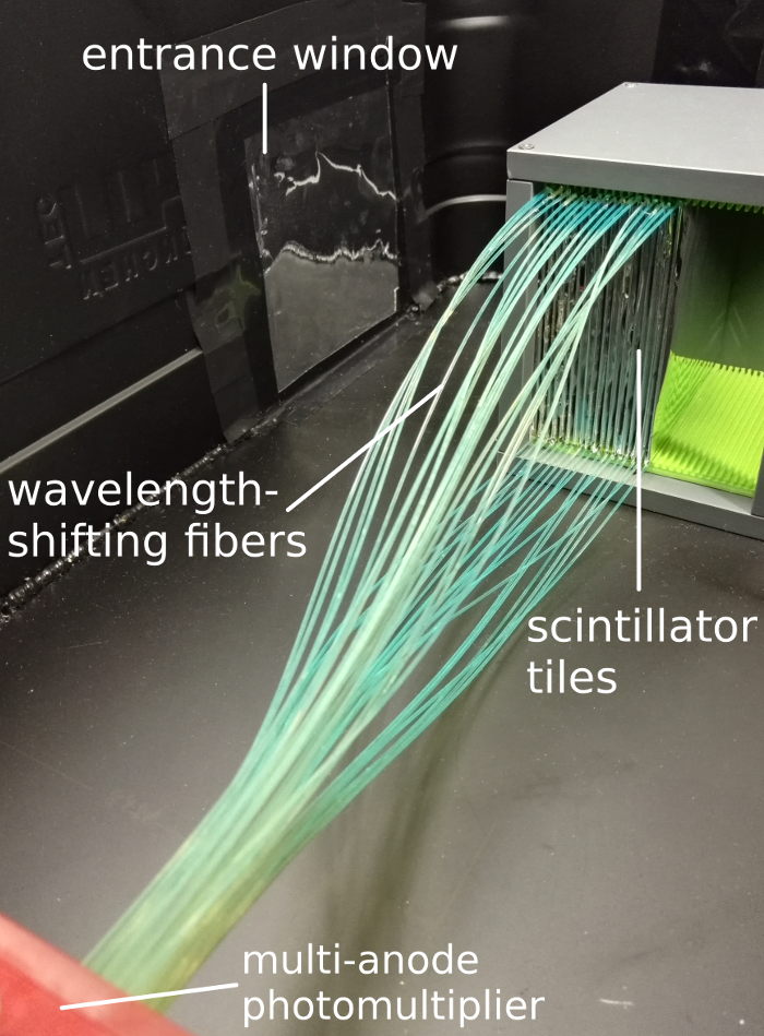
The contrast information in an ion range imaging system is determined from a measurement of the residual ion range behind the imaged object. It can be measured in a range detector, consisting of many thin scintillator planes. We are working on the improvement of a small prototype, consisting of 18 layers with 1mm thickness, read out via a multi-anode photomultiplier.
This detector combines simplicity with a fully active absorber material and thus promises excellent range detection capabilities.
In the context of the Compton-camera development several aspects of alternatives to the presently used design are being evaluated:
-
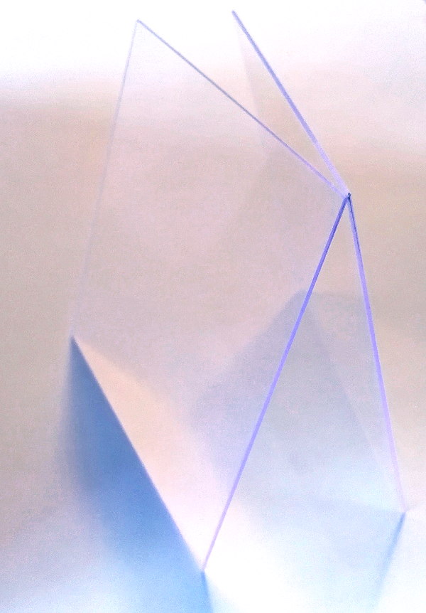
While presently LaBr3(Ce) is favored as scintillation material for the absorber component of the Compton camera, also CeBr3 is investigated as an alternative. It has the advantage of the absence of internal radioactivity (ca. 2 Bq/cm3 in case of LaBr3(Ce)) and the economic advantage of being (at present) less expensive while featuring almost the identical detector properties (energy and time resolution).
-
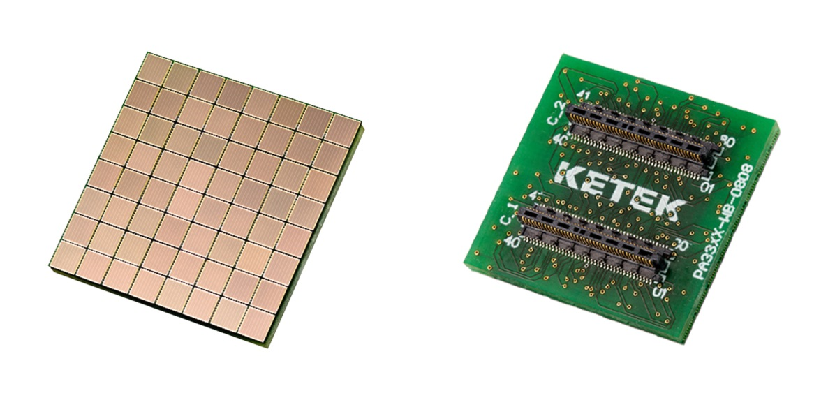
Position-sensitive readout of the monolithic absorber crystal is so far performed via segmented multi-anode photomultiplier tubes (MA-PMT). Starting with a high granularity of 16x16 segments (each ca. 3x3 mm2), we already learned that reducing the photosensor granularity to 8x8 segments would not result in a degradation of the spatial resolution (presently ca. 2.9 mm at 1.33 MeV photon energy). However, in the presence of magnetic fields (as for the scenario of PET/MR), a magnetic insensitive photosensor is required. Silicon Photomultipliers (SiPM) provide this capability and become increasingly popular as replacements of traditional PMTs. Together with the Munich-based company KETEK we are evaluating the performance of SiPM arrays, read out by an ASIC- (application specific integrated circuit) and FPGA-(field programmable gate array) based signal processing electronics. The photographs below show an 8x8 SiPM array from KETEK, as it will be characterized as photosensor of our Compton camera absorber detector.
-
Adding electron tracking capability to a Compton camera design is viable as long as the photon energies are high enough (ca. 1-2 MeV). For lower energies, like in nuclear medicine with (γ-)PET applications, an axially monolithic (yet laterally position-sensitive) scatter component has to be favored. Together with our Japanese collaborators from QST-NIRS (Chiba), we characterize a pixelated scintillator array from GAGG crystals (0.9x0.9x6) mm3.
Contact:
PD. Dr. P. Thirolf, Dr. J. Bortfeldt
References:
- S. Aldawood et al., Radiation Physics and Chemistry 140C (2017), 190-197.
- S. Liprandi et al., Conf. Record IEEE-NSS-MIC 2017.
Currently funded projects:
- 2018-2021, MultiSiP (together with KETEK GmbH), funded by Bayerische Forschungsstiftung, PI: P. Thirolf
- 2012-2018, Project C3.1, funded by DFG via Cluster of Excellence MAP (Munich-Centre for Advanced Photonics), PI: P. Thirolf, K. Parodi
- 2016-2020,MediNet: Network within the Integrating Initiative ENSAR2, funded by EU within Horizon 2020, PI: P. Thirolf